Mathematical Biology Seminar
Robert Marc
Moran Eye Center, University of Utah
Analyzing High Resolution Retinal Connectomes
Wednesday, April 27, 2016, at 3:05pm
LCB 219
 A connectome is a graph of cellular connectivity. What do we mean by connectivity? How do we define cells? What kind of graphs are these? How will we know if they are complete? We will explore these definitions as motivators for building connectomes rather than simply tracing networks.
We can build ultrastructural connectomes at high resolution with automated transmission electron microscope imaging, computational image assembly, and fusion of molecular markers with electron imaging. We will briefly review the physics, chemistry and biochemistry of connectome assembly. But once a connectome is built, we have accomplished essentially nothing.
Annotation is central to connectomics and is the most time-consuming stage. We cannot yet automate it. But once a connectome is richly annotated, we still have almost nothing.
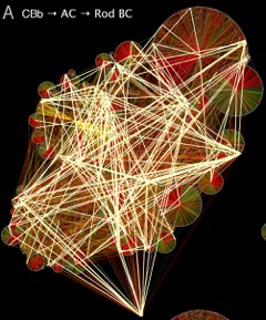
The whole point of building connectomes is the analysis of wiring. How does one explore an annotated volume? Searching sparse connectomes requires targeted hypotheses and directed tracing, but rich connectomes allow unbiased explorations using graphs. Our key example will be the discovery of new motifs in retinal organization that link night and day vision in mammals. These motifs allow rod and cone pathways to mutually inhibit each other in a winner-take-all architecture, which is central to most neural decision theories. But if they are so important, why weren't these motifs found earlier? We will explore how sparse but pervasive and powerful motifs can be hidden amidst the hoi polloi of common motifs and network hubs. Graph queries emerge as powerful hypothesis testing machines fed by connectomics.
A connectome is a graph of cellular connectivity. What do we mean by connectivity? How do we define cells? What kind of graphs are these? How will we know if they are complete? We will explore these definitions as motivators for building connectomes rather than simply tracing networks.
We can build ultrastructural connectomes at high resolution with automated transmission electron microscope imaging, computational image assembly, and fusion of molecular markers with electron imaging. We will briefly review the physics, chemistry and biochemistry of connectome assembly. But once a connectome is built, we have accomplished essentially nothing.
Annotation is central to connectomics and is the most time-consuming stage. We cannot yet automate it. But once a connectome is richly annotated, we still have almost nothing.
The whole point of building connectomes is the analysis of wiring. How does one explore an annotated volume? Searching sparse connectomes requires targeted hypotheses and directed tracing, but rich connectomes allow unbiased explorations using graphs. Our key example will be the discovery of new motifs in retinal organization that link night and day vision in mammals. These motifs allow rod and cone pathways to mutually inhibit each other in a winner-take-all architecture, which is central to most neural decision theories. But if they are so important, why weren't these motifs found earlier? We will explore how sparse but pervasive and powerful motifs can be hidden amidst the hoi polloi of common motifs and network hubs. Graph queries emerge as powerful hypothesis testing machines fed by connectomics.